
Guided dental implant surgery begins long before the procedure itself. This article explains how CBCT imaging, digital scans, thoughtful treatment planning, and custom surgical guides work together to improve the accuracy, predictability, and long-term success of dental implant treatment.
Key takeaways
- Most of the work behind a guided implant happens during planning, before you sit in the treatment chair.
- A CBCT scan shows the bone and nerves in three dimensions. A digital scan captures your teeth, gums, and bite.
- The implant is planned around the ideal final crown, and a custom guide transfers that plan to surgery.
- The guide is checked for a complete, accurate fit before any implant is placed.
Why Guided Dental Implant Surgery Begins Before the Procedure
When most people think about getting a dental implant, they picture the titanium implant that replaces the root of a missing tooth or the final crown they’ll eventually see when they smile. Very few realize that one of the most important parts of the entire procedure is never permanently placed in the mouth—and is used for only a short time during surgery.
It’s called a surgical guide.
I’ve been placing dental implants since 1999, and one of the biggest changes I’ve seen is what happens long before the procedure itself. Today, much of the work that influences the success of an implant takes place well before a patient sits in the treatment chair.
To most patients, a surgical guide looks like nothing more than a simple piece of clear plastic. To me, it represents hours of diagnosis, digital planning, and clinical judgment, all directed toward placing your implant in its ideal position while respecting your unique anatomy, available bone, and restorative goals.
That preparation helps reduce uncertainty during surgery and creates a stronger foundation for a healthy, functional, and natural-looking final crown.
The guide itself isn’t what makes an implant successful. The planning behind the guide is what makes the difference.
In many ways, implant surgery begins with the end in mind. Before I decide where an implant should go, I first determine where your new tooth should ideally be positioned for appearance, function, and long-term health.
From there, I work backward to determine the implant position that best supports that tooth while maximizing healthy bone support, preserving bone and gum tissue whenever possible, and avoiding important structures such as adjacent tooth roots, nerves, and the maxillary sinus.
One of the goals of digital planning is to answer as many important questions as possible before the day of surgery. By the time your procedure begins, the critical decisions have already been made. Surgery then becomes the careful execution of a well-developed treatment plan rather than the time to make those decisions.
That often makes surgery more efficient, reduces variables during the procedure, and, in many cases, allows me to use a minimally invasive approach. Instead of making an incision and lifting the gum tissue away from the bone, the implant can often be placed through a small opening in the gum. This typically results in less swelling, less discomfort, and a quicker recovery.
When patients ask why I use guided implant surgery, the answer isn’t because I like technology.
It’s because I believe the best implant treatment starts with a carefully developed plan—one that allows surgery to be performed with greater precision, confidence, and consistency while being tailored to each patient’s unique anatomy.
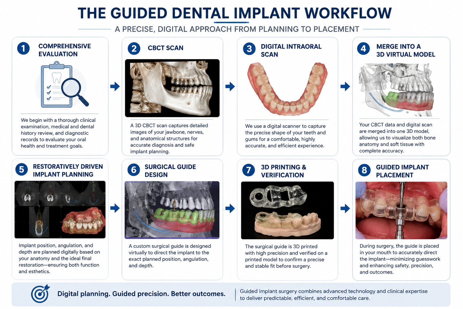
How Implant Dentistry Has Evolved Since 1999
When I placed my first dental implant in 1999, implant dentistry looked very different than it does today. We relied primarily on two-dimensional X-rays, stone models, surgical stents, and clinical experience to determine implant position. Those techniques produced many successful, long-lasting implants, but they required considerably more interpretation than the digital planning tools available today.
Then
- Two-dimensional X-rays
- Conventional impressions and stone models
- Relatively simple positioning templates
Today
- Three-dimensional CBCT imaging
- Digital intraoral scans
- Precise, computer-planned surgical guides
Over the past 25 years, I’ve worked with numerous implant systems and have watched nearly every aspect of implant dentistry evolve. Implant designs, surface treatments, restorative components, digital imaging, planning software, and surgical techniques have all advanced dramatically. Each advancement has improved the precision, consistency, and predictability of implant treatment.
One of the most significant advances has been the ability to evaluate and plan implant treatment in three dimensions before the procedure ever begins.
Three-dimensional CBCT imaging has transformed how I evaluate bone and surrounding anatomy. Digital intraoral scanners have replaced many conventional impressions. Modern planning software allows me to evaluate the relationship between your bone, surrounding teeth, gum tissue, bite, and the planned final crown long before an implant is ever placed.
Surgical guides have evolved as well. What were once relatively simple positioning templates have become highly precise guides that integrate CBCT imaging, digital scans, and computer-guided planning. They allow a digital treatment plan to be transferred accurately and consistently from the computer to your mouth in a way that simply wasn’t possible when I began placing implants.
Although digital technology has transformed implant dentistry over the past 25 years, one thing has remained constant: technology alone doesn’t make implant treatment successful.
Throughout my career, I’ve adopted new technology only when it clearly improves patient care, enhances long-term outcomes, or allows treatment to be performed more consistently. Experience, sound clinical judgment, and careful diagnosis remain far more important than any individual piece of technology.
Guided implant surgery isn’t about the technology—it’s about making better decisions before surgery begins. Modern digital tools allow me to better understand your anatomy, identify potential challenges before surgery, and develop a treatment plan tailored specifically to you. That’s why every implant I place is digitally planned and placed using a custom surgical guide—not because freehand implant placement cannot be successful, but because I believe this workflow is the most dependable way to carry out a carefully developed treatment plan.
Understanding Your Jaw in Three Dimensions
Successful implant treatment begins with understanding your unique anatomy. While traditional dental X-rays provide valuable diagnostic information, they can show only a two-dimensional view of structures that exist in three dimensions. For implant planning, I need a more complete picture.
To gain that understanding, I use Cone Beam Computed Tomography (CBCT), a specialized three-dimensional scan that allows me to evaluate your jaw from virtually every angle before treatment begins. Rather than estimating what lies beneath the gums, I can evaluate your anatomy in far greater detail.
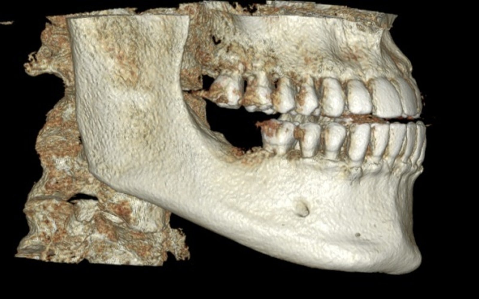
The CBCT allows me to evaluate the quantity and quality of your available bone—including its width, height, shape, and density—before deciding where an implant should be placed. It also reveals important anatomical structures such as the inferior alveolar nerve, the maxillary sinuses, neighboring tooth roots, and other landmarks that must be carefully considered during treatment planning.
One of the greatest advantages of CBCT imaging is that it helps answer important questions before the day of surgery. Instead of discovering potential challenges during the procedure, I can identify them during the planning process, giving me the opportunity to choose the treatment approach that’s most appropriate for you.
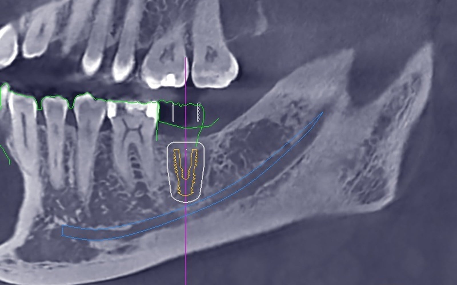
For example, having enough bone to place an implant doesn’t necessarily mean it’s the ideal location. The available bone, planned position of the final crown, surrounding gum tissue, bite, and nearby anatomical structures all have to work together. The best implant position is the one that balances all of these factors—not simply the first place an implant will fit.
Sometimes that means recommending bone grafting before implant placement. Other times it may involve selecting a different implant diameter or length, adjusting the implant position or angulation, or even recommending a different treatment approach altogether. Every recommendation is based on your individual anatomy and treatment goals rather than a one-size-fits-all philosophy.
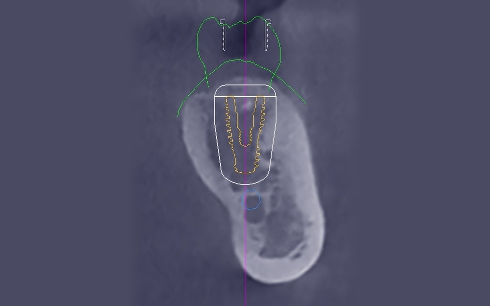
The goal isn’t simply to find a place where an implant will fit. The goal is to identify the position that provides the best balance of stability, function, esthetics, healthy surrounding tissues, and long-term maintainability.
Bone is only one part of successful implant planning. Next, I combine this information with a highly accurate digital scan of your teeth and gums, allowing me to see both the anatomy beneath the gums and the teeth and gums above them within a single virtual model.
Combining Your Bone, Teeth, and Gums into One Virtual Model
While the CBCT provides exceptional detail about the bone and structures beneath the gums, it cannot capture the exact shape of your teeth, your bite, or the contours of your gum tissue. Those details are equally important because the implant must ultimately support a crown that looks natural, functions comfortably, and remains healthy for many years.
To capture that information, I use an intraoral scanner to create a precise three-dimensional digital model of your teeth and gums. Unlike traditional impressions, which rely on impression material and stone models, digital scanning records the visible surfaces of your mouth with exceptional precision while providing a more comfortable experience for most patients.
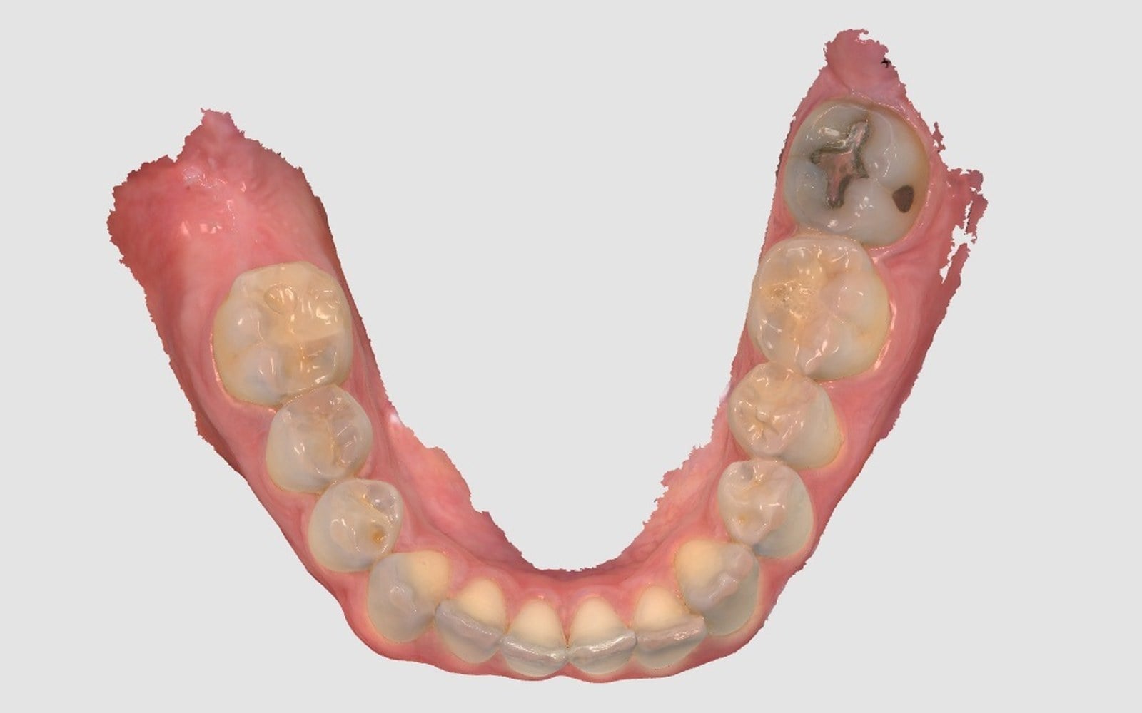
The digital scan allows me to evaluate how your teeth fit together, the contours of your gums, the available space for the final crown, and how it will function within your bite.
The real value isn’t the CBCT scan or the digital scan individually—it’s how they work together.
Specialized planning software merges these two sources of information into a single comprehensive virtual model of your mouth. Instead of evaluating the bone, teeth, and gums separately, I can see how they all relate to one another while planning your implant.
This combined virtual model allows me to evaluate every aspect of your treatment before surgery begins. Rather than mentally piecing together information from multiple sources, I can evaluate everything within one integrated model.
The virtual model also allows me to show you exactly what I see. Instead of simply explaining why I recommend a particular treatment, I can review your anatomy with you, discuss your treatment options, and answer your questions using your own three-dimensional images. I believe patients make better decisions when they understand both the problem and the proposed solution.
Perhaps the greatest advantage of digital planning is that it allows me to plan the implant around the final crown—not force the final crown to adapt to the implant.
Before deciding where an implant should be placed, I first determine where the final crown should ideally be positioned for appearance, function, long-term health, and ease of maintenance. I then work backward to determine the implant position, angulation, depth, and size that best support that goal while respecting your available bone and surrounding anatomy.
This philosophy is known as restoratively driven implant planning, and it has become one of the guiding principles of modern implant dentistry.
Rather than asking, “Where can I place an implant?” the better question is, “Where should the new tooth be?”
Whether replacing a front tooth or a back tooth, planning the final crown first leads to better implant placement. For front teeth, this helps create natural-looking gum contours, proper symmetry, and an attractive smile. For back teeth, it helps distribute biting forces appropriately while making the restoration easier to clean and maintain for years to come.
Once the treatment plan is complete, the surgical guide is designed to accurately transfer those decisions during your procedure. Rather than making important decisions during surgery, my focus becomes carrying out a treatment plan that has already been carefully developed.
Successful implant treatment doesn’t begin with placing an implant. It begins with understanding the complete picture and developing the right plan. The surgical guide simply allows that plan to be carried out exactly as intended.
Planning the Ideal Implant Position
Most people assume that placing a dental implant is simply a matter of finding enough bone to hold it securely. In reality, that’s only one piece of the puzzle.
Every successful implant requires balancing multiple factors. The ideal position of the final crown, the available bone, your gum tissue, bite, nearby teeth, important anatomical structures, and your ability to keep the implant clean all influence where the implant should ultimately be placed.
Balancing these factors helps create an implant that looks natural, functions comfortably, remains healthy, and serves you well for decades.
Although the planning principles remain consistent, every implant treatment plan is customized to your anatomy, oral health, and long-term goals.
Successful implant treatment is rarely determined by a single decision. Instead, it reflects the cumulative effect of many well-informed decisions.
Four of the most important planning decisions involve the implant’s position, angulation, depth, and size. Although these may sound like technical details, each one directly influences how your implant will look, function, and perform over the long term.
Position
The position of an implant determines far more than where it sits in the bone. It influences how natural the final crown will look, how the surrounding gums are supported, how biting forces are distributed, and how easily the implant can be cleaned for years to come.
The ideal implant position supports both the final crown and the long-term health of the surrounding tissues.
Angulation
The angle of an implant is equally important. Proper angulation helps the final crown function naturally while directing chewing forces through the implant in the way it was designed to handle them.
Whenever possible, I plan implants to support screw-retained crowns. These restorations eliminate the need for cement beneath the gums, are easier to remove if maintenance is ever needed, and often provide greater flexibility throughout the life of the implant.
Depth
Depth is another important consideration. Placing the implant at the proper depth helps create a natural emergence profile—the way the final crown appears to emerge from the gums—while supporting healthy gum tissue and preserving the surrounding bone.
Proper implant depth contributes to both the appearance of the restoration and its long-term health.
Size
Selecting the appropriate implant diameter and length is equally important. Rather than automatically selecting the largest implant available, I choose the size that best fits your anatomy while providing excellent stability and long-term support.
Selecting the appropriate implant size helps preserve healthy bone, respect nearby anatomical structures, and provide a strong foundation for the final crown.
Every planning decision influences the others. A small adjustment in implant position may affect angulation, depth, implant size, or the final restoration, which is why successful implant planning is about finding the right balance rather than optimizing a single measurement.
You don’t need to understand every technical detail of implant planning. My goal is simply to help you understand why these planning decisions play such an important role in achieving a predictable, long-lasting result.
No single planning decision determines success. Long-term results are built on many well-informed decisions made before surgery ever begins.
Designing the Surgical Guide
Once the digital treatment plan is complete, the next step is designing the custom surgical guide that will be used during your implant procedure.
Every surgical guide is custom designed for a specific patient and procedure. It is not an off-the-shelf device. Instead, it is designed using your CBCT scan and digital scan to fit your mouth precisely and transfer your personalized treatment plan during surgery.
Designing the guide requires combining two types of digital information. Each provides different information, and both are essential to the final design.
The digital scan captures the exact shape of your teeth and gums. This surface detail allows me to design a guide that fits securely and consistently throughout the procedure.
The CBCT scan reveals the bone and important anatomical structures beneath the surface. It allows me to determine the ideal implant position, angulation, depth, and size. The planning software then positions the guide sleeve so the surgical instruments follow the exact path established during planning.
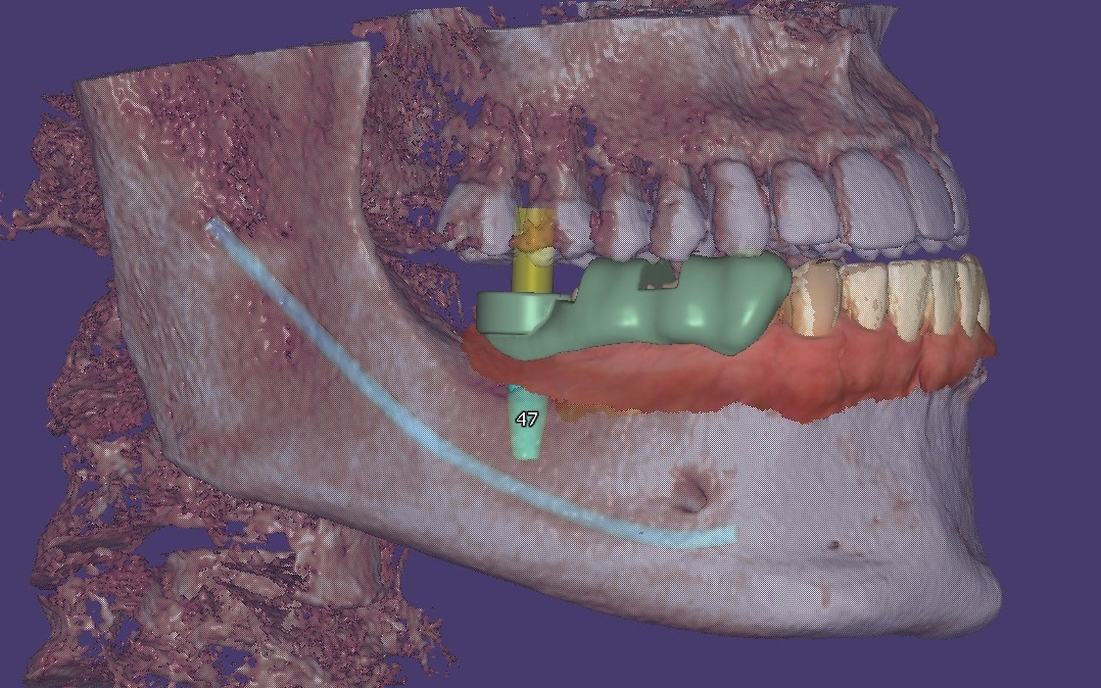
Together, these two scans allow the surgical guide to accurately transfer your treatment plan during surgery. By the time the guide is complete, the key treatment decisions have already been made, allowing the procedure to focus on carrying out those decisions rather than making them during surgery.
Although the surgical guide appears simple, every contour, opening, and guide sleeve has been intentionally designed for your specific treatment. Each feature reflects careful diagnosis, digital planning, and clinical judgment.
Before your procedure begins, I always verify that the guide seats completely and accurately on your teeth. Even though it has been digitally designed, confirming that it fits exactly as planned is an important step in providing safe, precise, and predictable treatment.
The value of the surgical guide isn’t the guide itself—it’s its ability to faithfully transfer your treatment plan to your mouth.
Once the design is complete, the guide is manufactured, inspected, and prepared for your procedure.
From Surgical Guide to Implant Placement
Once the design is complete, your custom surgical guide is 3D printed using a professional dental printer and a biocompatible dental resin.
After printing, the guide is carefully cleaned, fully cured, inspected, and prepared for clinical use. A guide sleeve is then securely incorporated to direct the surgical instruments exactly as planned.
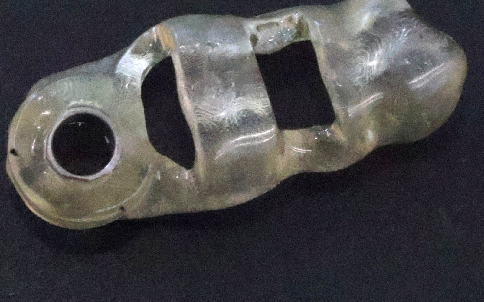
Before implant placement begins, I verify that the guide seats completely and accurately on your teeth. Although the guide has been carefully designed and manufactured, I never assume everything is perfect. Confirming that it fits exactly as intended is an essential quality check before the procedure begins.
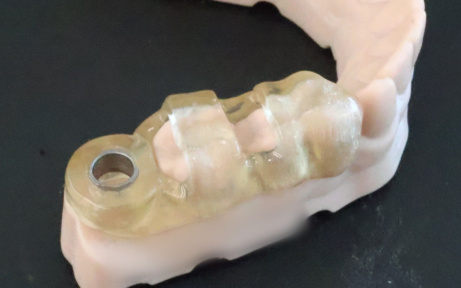
Only after confirming the fit do I begin implant placement.
By the time surgery begins, every important treatment decision has already been made. My focus shifts from planning the procedure to carefully carrying it out.
The surgical guide translates the digital treatment plan into the clinical procedure, helping implant placement closely follow the planned position, angulation, depth, and size.
Successful guided implant surgery reflects the entire treatment process—from accurate diagnosis and digital treatment planning to custom guide design, 3D printing, clinical verification, and meticulous surgical execution. Each step contributes to a more consistent procedure and supports a predictable, long-lasting result.
Conclusion
By the time your surgical guide is used during implant placement, the important clinical decisions have already been made. That guide represents the final result of a carefully coordinated process that begins long before the day of surgery.
Throughout this article, I’ve explained how CBCT imaging, digital scanning, restorative-driven implant planning, and custom surgical guides work together to support accurate, predictable dental implant placement.
Technology has transformed implant dentistry, but it hasn’t replaced the principles of successful treatment. Experience, clinical judgment, and personalized treatment planning remain the foundation of every implant I place. Digital technology simply allows me to apply those principles with greater consistency.
My goal has never been to use technology for its own sake. It’s to use every available tool thoughtfully to better understand your anatomy, plan your treatment with confidence, and place your implant as accurately as possible. When that process is guided by experience and sound clinical judgment, it creates the best opportunity for an implant that looks natural, functions comfortably, remains healthy, and serves you well for many years.
Frequently Asked Questions About Guided Dental Implant Surgery
About Guided Surgery
What is guided dental implant surgery?
Guided dental implant surgery is a technique that combines CBCT imaging, digital scans, specialized planning software, and a custom surgical guide to plan implant placement before surgery begins. Rather than making important treatment decisions during the procedure, those decisions are made in advance using detailed information about your anatomy. The surgical guide then helps carry that plan out during implant placement.
Does every dental implant require a surgical guide?
Not necessarily. Experienced implant dentists can successfully place implants freehand in many situations. In my practice, however, every implant is digitally planned and placed using a custom surgical guide because I believe it provides the most consistent way to carry out treatment while respecting each patient’s unique anatomy.
Are surgical guides more accurate than freehand implant placement?
Clinical studies have shown that guided implant surgery can improve the accuracy and consistency of implant placement. However, the guide is only one part of the process. Careful diagnosis, personalized treatment planning, and clinical judgment remain equally important to achieving an excellent long-term result.
Planning Your Implant
Why is a CBCT scan necessary before placing a dental implant?
Traditional dental X-rays provide only a two-dimensional view of your teeth and jaws. A CBCT scan creates a detailed three-dimensional image that allows me to evaluate your bone, nearby nerves, tooth roots, sinuses, and other important anatomical structures before treatment begins. This information helps me develop a treatment plan that is tailored to your individual anatomy.
How long does implant planning take?
Planning often takes longer than the implant surgery itself. I carefully review your CBCT scan, evaluate your anatomy, determine the ideal implant position, design your surgical guide, and verify every aspect of the treatment before your appointment. Investing more time in planning helps create a smoother and more predictable procedure.
Will I be able to see my treatment plan before surgery?
Yes. One advantage of digital planning is that I can review your CBCT scan, digital models, and proposed implant position with you before treatment begins. Many patients find it easier to understand their options when they can see their own anatomy and discuss the plan together.
What if I don’t have enough bone for a dental implant?
Not having enough bone doesn’t automatically mean you cannot receive an implant. Depending on your situation, treatment may include bone grafting, selecting a different implant, modifying the implant position, or considering another treatment approach. A CBCT scan allows these options to be evaluated before surgery.
During Treatment
Can a dental implant be placed immediately after a tooth is removed?
Often, yes. In many situations, an implant can be placed during the same appointment as the extraction. Whether this is appropriate depends on factors such as infection, available bone, implant stability, and your overall treatment goals.
Can guided surgery be used for multiple implants or full-mouth implant reconstruction?
Yes. Guided surgery can be used for single implants, multiple implants, implant-supported bridges, and full-arch implant reconstruction. As treatment becomes more complex, digital planning often becomes even more valuable because multiple implants must work together to support the final restoration.
Does guided implant surgery reduce discomfort and recovery time?
In many cases, guided planning allows implants to be placed through smaller incisions or, in some situations, without raising a large surgical flap. This may result in less swelling, less discomfort, and a faster recovery. However, every patient and every procedure is different, so treatment is always tailored to your individual needs.
What happens if the surgical guide doesn’t fit properly?
Before implant placement begins, I always verify that the guide fits completely and accurately. If it does not fit exactly as expected, I determine the cause before proceeding. Patient safety and accurate implant placement always take priority over following the original plan.
Long-Term Success
Does guided implant surgery guarantee success?
No. No technology or surgical technique can guarantee a successful outcome. Guided surgery improves planning and helps accurately carry out that plan, but long-term success also depends on healing, bone quality, oral hygiene, general health, smoking, regular professional maintenance, and ongoing home care.
Is every implant treatment plan customized?
Yes. Every patient has different anatomy, bone volume, gum contours, bite relationships, medical considerations, and treatment goals. While the planning process follows the same principles, every implant treatment plan is personalized to the individual patient.
Why doesn’t every implant dentist use guided surgery?
Guided implant surgery requires additional imaging, digital planning software, laboratory workflows, and training. Many implant dentists routinely use guided surgery, while others may prefer freehand techniques depending on the clinical situation. In my practice, I digitally plan every implant because I believe it provides the most consistent way to deliver individualized treatment.
What should I look for when choosing an implant dentist?
There isn’t a single credential or piece of technology that guarantees excellent implant care. Ask how your anatomy will be evaluated, whether three-dimensional imaging is used when appropriate, how treatment is planned before surgery, what experience the dentist has with cases similar to yours, and how long-term maintenance will be managed. Technology is an important tool, but successful implant treatment ultimately depends on accurate diagnosis, personalized treatment planning, sound surgical technique, and clinical experience.
References and Further Reading
The information in this article is based on current scientific literature, published clinical guidelines, and the author’s clinical experience. The references below provide additional information for readers interested in learning more about guided implant surgery and modern implant dentistry.
- American Academy of Implant Dentistry. Dental Implants. Available from the AAID website.
- American Dental Association. Dental Implants. ADA Oral Health Topics.
- International Team for Implantology. ITI Treatment Guide Series.
- European Association for Osseointegration. Clinical Guidelines and Consensus Statements.
- Per-Ingvar Brånemark, Zarb GA, Albrektsson T. Tissue-Integrated Prostheses: Osseointegration in Clinical Dentistry. Quintessence Publishing.
- Jung RE, Zembic A, Pjetursson BE, Zwahlen M, Thoma DS. Systematic review of the survival rate and complications of implant-supported single crowns. Clinical Oral Implants Research.
- Pjetursson BE, Asgeirsson AG, Zwahlen M, Sailer I. Improvements in implant dentistry over time: systematic reviews and long-term outcome studies. Clinical Oral Implants Research.
- Morton D, Gallucci G, Lin WS, et al. Group 2 ITI Consensus Report: Prosthodontics and implant dentistry. Clinical Oral Implants Research.
- Tahmaseb A, Wismeijer D, Coucke W, Derksen W. Computer-guided implant placement: a systematic review of accuracy and clinical outcomes. International Journal of Oral & Maxillofacial Implants.
- Block MS. Color Atlas of Dental Implant Surgery. Elsevier.
- Misch CE. Dental Implant Prosthetics. Elsevier.
- Buser D, Sennerby L, De Bruyn H. Modern implant treatment concepts and long-term evidence. Clinical Oral Implants Research.
Medical Disclaimer: This article is intended for educational purposes only and should not replace a comprehensive dental examination or personalized treatment recommendations. Every patient is unique, and treatment decisions should be based on an individual clinical evaluation.


